BIO 102 MENU
syllabus 
1 - origin
2 - biomol.
3 - biomol2
4 - viruses
5 - prokaryon
6 - endosym
7 - eukaryon
8 - energy
9 - mitosis
10 - meiosis
11 - reprod
12 - genetics
13 - humgene
14 - humge2
15 - evolution
16 - evolutio2
17 - diversity
18 - diversi2
19 - tissues
20 -digestive
21 - respirat
22 - circul
23 - excret
24 - endocr
25 - receptors
26 - nervsys
Quizzes
Bio 103 Lab
(full title of lecture appears in status bar on the top or at the bottom of your window)

Biology 102 - General Biology
Animal Structure and Function
Coordinating System: The Nervous System
Receptors-->Sensory neurons (PNS)-->Interneurons (CNS)-->Motor neurons (PNS)-->Effectors
The Nervous System
Vertebrates all have a dorsal hollow nerve cord. It forms on the dorsal side of the embryo as the neural tube. The brain and the spinal cord derive from the neural tube. The neural tube closes early in embryogenesis and failure to close can result in a baby who is anencephalic (without a brain) or who has spina bifida (an opening somewhere along the spine). The child is paralyzed from the opening down. Folate or folic acid, one of the B vitamins, is known to reduce the risk of such an anomaly. Folate is also good for your heart and everyone is advised to take it. However, women of childbearing age should be taking it all the time or eating foods that are rich in it. If a person has already had a child with a neural tube defect (NTD) the mother is advised to take 4 mg/day of folate (or folic acid) at least three months before and three months after conception in future pregnancies. Hispanics have a higher incidence of NTDs than other ethnic groups.
The nervous system is far superior in speed and selectivity to the endocrine system. It depends on a specialized system of nerve cells called neurons which receive and give instructions by means of electrical impulses directed over specific pathways. The neurons are highly differentiated cells and are similar in all animals. Their structure and function are intimately related. The resting neuron (like all cells) has a charge difference across its membranes due to the differential distribution of ions (thanks to the Na+ /K+ pump in cell membrane). When a neuron is stimulated, the resting potential gives way to the action potential and the membrane is depolarized. The depolarization travels in one direction down the axon of the neuron to the opposite end of the cell. At the "far" end, the neuron has small vesicles containing a neurotransmitter which is released when the membrane depolarization occurs there. When the neurotransmitter is released, it binds to receptors in the membrane of the next cell (either another neuron or an effector cell), and depolarizes it. If the cell is another neuron, the depolarization begins in that cell and travels along its axon. If the next cell is a muscle, the muscle cell membrane is depolarized (in a manner similar to the neuron) and a series of reactions occur within the muscle resulting in either contraction or relaxation of the muscle.
Neurotransmitters can be excitatory or inhibitory. Each muscle is enervated by both kinds. Endocrine glands or exocrine glands, the other kind of effector cells or organs, respond by releasing their product when stimulated by a motor neuron. It is interesting to note that even in the highly efficient nervous system, the ultimate messenger between cells is a chemical. So even here we see the reliance on a process very similar to the evolutionary older system of hormonal (chemical) communication!
CENTRAL NERVOUS SYSTEM CNS): BRAIN AND SPINAL CORD NEURONS
The nervous system is anatomically divided into the central nervous system (CNS) and the peripheral nervous system (PNS). The CNS consists of the brain and spinal cord which contain inter neurons. The neurons of the CNS are referred to as inter neurons. They make connections with many other neurons including neurons within the CNS as well as the sensory and motor neurons of the PNS. The neurons of the CNS send messages to many effector cells and receive messages from many receptor cells. It is their job to integrate the information received and coordinate the body's responses.

THE PERIPHERAL NERVOUS SYSTEM (PNS): THE CRANIAL NERVES AND THE SPINAL NERVES
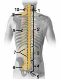
The PNS is composed of the sensory neurons and the motor neurons that come respectively from the receptors and go to the effectors. There are 12 cranial nerves connected to the brain. Some are sensory, some motor and some mixed. There are 31 pairs of spinal nerves which are connected to the spinal column. They are both sensory and motor nerves. The word, nerve, refers to a collection of neurons.
 z
z
THE 12 CRANIAL NERVES
The cranial nerves are part of the peripheral nervous system.
Some are composed of sensory neurons, some of motor neurons and some are mixed.
The (motor) neurons of the peripheral nervous system are divided functionally into the somatic and autonomic subdivisions. The somatic subdivision refers to those cranial and spinal nerves that carry messages to voluntary skeletal muscles. The autonomic nervous system is that part of the peripheral nervous system that is not under voluntary control. The autonomic nerves are those cranial and spinal nerves that carry messages to the smooth muscle of the internal organs: uterus, stomach, heart, etc., and most glands. In the voluntary system, the motor neurons originate in the spinal cord and run uninterrupted to the effector. In the autonomic, an additional motor neuron is inserted.
An additional functional separation exists within the autonomic system. It consists of the sympathetic and parasympathetic motor nerves. All organs are innervated by both types of nerves. The sympathetic and parasympathetic work antagonistically but result in fine tuning the body needs. At times one takes precedence over the other.

The parasympathetic consists of some of the cranial nerves from the brain (e.g., the vagus nerve, 10th cranial nerve) and some from the sacral region of the spinal cord. Acetylcholine is the chemical transmitter at the parasympathetic synapses. Its function is to stimulate digestion, slow the heart and lower the blood supply to skeletal muscles. The parasympathetic signals a "vegetative" state. After a full meal, the parasympathetic takes over and the blood is diverted to the digestive tract and away from the skeletal muscles and brain.
The sympathetic consists of nerves from the thoracic and lumbar regions of the spinal cord and the nerves form a chain of ganglia immediately outside the vertebral column, "the sympathetic chain," composed of cell bodies (nuclei) of additional motor neurons. The parasympathetic system also has ganglia but they are close to the target organ and far from the CNS. When the sympathetic signals are predominating, the body assumes a "fight or flight" response. If something scares you, the sympathetic takes over and causes a release of adrenalin (epinephrine) so that the blood goes to the skeletal muscles and heart and away from the digestive tract. The sympathetic system also stimulates the adrenal gland, thus even more adrenalin (epinephrine) is released to help the body fight or flee. Epinephrine is both a neurotransmitter and a hormone.
When you take the final in this class, do not eat too much beforehand or your blood will go to your stomach and not to your brain! As you read the exam the photoreceptors in your retina will be stimulating the sensory neurons of your optic nerve (a cranial nerve which is part of your PNS). The optic nerve endings will stimulate the inter neurons of your brain which in turn will stimulate others (we hope) to remember the answers to the questions. When you have the answer, the inter neurons of the brain and spinal cord will stimulate the motor neurons in the somatic subdivision of your PNS and they, in turn, will stimulate the muscles (effector cells) of your arm and fingers to write the right answers!! Whew!! Good luck.